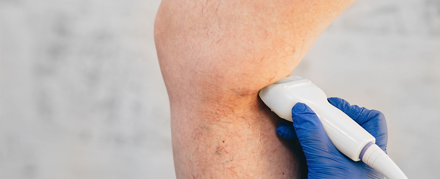
Ultrasound Evaluation
Venous Duplex Ultrasound is the gold standard for diagnosis of vein disease. At CVC, the duplex ultrasound is usually done at your initial consultation visit by a qualified, venous ultrasound technician. The technicians look especially at the leg’s major superficial veins, the great saphenous vein and the small saphenous veins, which lie under the surface of the skin. These veins, when abnormal and refluxing, are usually the root of most Varicose veins medical problems. The ultrasound evaluation provides many valuable pieces of information about the leg veins and is vital in establishing an accurate diagnosis, which is the only way to assure the most effective treatment of your varicose veins.
First, it determines if there is healthy flow or unhealthy reflux in the veins. The ultrasound image provides information about the blood flow inside the leg veins and can determine if the vein has normal, healthy valves that work against gravity taking blood from the legs back to the heart. It can also determine if the vein has unhealthy valves allowing blood to flow backwards, or reflux, toward the feet.
Second, the ultrasound can determine the size, location and the various branches and connections are “mapped-out.” This enables a record to be made showing the precise location of the leaking valves.
Third, the ultrasonographer can also evaluate your leg’s deep vein system for the presence of a dangerous blockage by a clot, also called a deep vein thrombosis (DVT).
